Marquette professor awarded $1.4 million grant to study metal reduction technique in CT imaging
“Oftentimes, metal implants can negatively alter CT images, making diagnosis challenging. Since CT scans are used to plan radiation therapy, these image degradations can lead to treatment inaccuracies.”
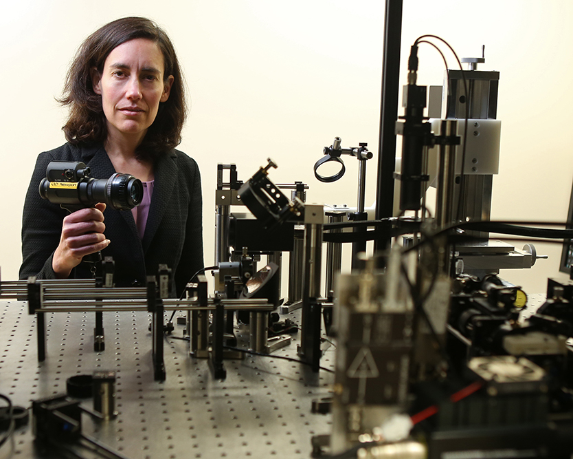
Taly Gilat-Schmidt. Photo courtesy of Marquette University.
MILWAUKEE — Dr. Taly Gilat-Schmidt, associate professor of biomedical engineering at Marquette University and the Medical College of Wisconsin, has received a $1.4 million grant from the National Institutes of Health to study metal artifact reduction techniques to improve the accuracy of computed tomography scans and radiation treatment.
Gilat-Schmidt and collaborator Dr. Emil Sidky of the University of Chicago will use the $1,405,211 RO1 grant to develop a method of reducing inaccuracies in CT imaging caused by metal within the body — such as implants and orthopedic hardware — which can obscure images of organs and tissues.
“Oftentimes, metal implants can negatively alter CT images, making diagnosis challenging. Since CT scans are used to plan radiation therapy, these image degradations can lead to treatment inaccuracies,” Gilat-Schmidt said. “By using a validated algorithm, our work aims to reduce this variability to help doctors better diagnose and plan radiation treatment for patients.”
Dr. Kristina Ropella, Opus Dean of the Opus College of Engineering, said she is thrilled for Gilat-Schmidt and her team, which includes Sidky and others from the University of Chicago, as well as the Department of Radiation Oncology at MCW.
“Her imaging research continues to draw well-deserved recognition among her peers and most importantly, her work is benefiting patient care outcomes,” Ropella said.
NIH is the largest public funder of biomedical research in the world, investing more than $32 billion annually to enhance life and reduce illness and disability.
About Gilat-Schmidt’s Medical Imaging Systems Laboratory
The Medical Imaging Systems Laboratory focuses on the design and optimization of medical imaging systems and reconstruction algorithms, with the goal of improving image quality and reducing radiation dosage. Application of theoretical, computational and experimental methods are used on CT, tomosynthesis and X-ray imaging. Laboratory collaborators include the Medical College of Wisconsin, University of Chicago and industry partners.
NOTE: This press release was submitted to Urban Milwaukee and was not written by an Urban Milwaukee writer. While it is believed to be reliable, Urban Milwaukee does not guarantee its accuracy or completeness.
Mentioned in This Press Release
Recent Press Releases by Marquette University

New Marquette Law School Poll finds large majority of Wisconsin voters not yet tuned in to who is running in major 2026 elections
Oct 29th, 2025 by Marquette UniversityNo candidate has established strong position in public favorability in governor, state Supreme Court races; large majorities of voters undecided